Información legal:
Titular de la página web: Gris Reproducción SL
Propiedad Intelectual:
Todos los derechos de explotación están reservados. Queda prohibida la reproducción, retransmisión, copia, ce-sión o difusión total o parcial de su contenido sin autorización expresa y por escrito. Tampoco se autoriza:
- La PRESENTATION de una página del sitio web en una ventana que no pertenezca a Gris Reproducción SL por medio de "framing".
- La inserción de una imagen difundida en el sitio web en una página que no sea de Gris Reproducción SL mediante "in line linking".
- La extracción de elementos del sitio web causando perjuicio a Gris Reproducción SL, de conformidad con las disposiciones vigentes.
Garantías y Responsabilidades:
Gris Reproducción SL no asume la responsabilidad de las infracciones en que puedan incurrir los usuarios ni de los daños y perjuicios causados por la utilización de esta web, y se reserva el derecho de actualización y modificación de la información sin previo aviso.
Gris Reproducción SL no asume la responsabilidad del contenido, la veracidad y los errores en los enlaces a los que se puede acceder a través de la página web. La única finalidad de los enlaces consiste en proporcionar a los usuarios el acceso a una información que pueda ser de su interés.
Las anteriores condiciones se regirán por la normativa española.
08017 - Barcelona - Spain
Telephone: (+34) 93 280 65 35
Fax: (+34) 93 280 08 71
doctorgris@doctorgris.com

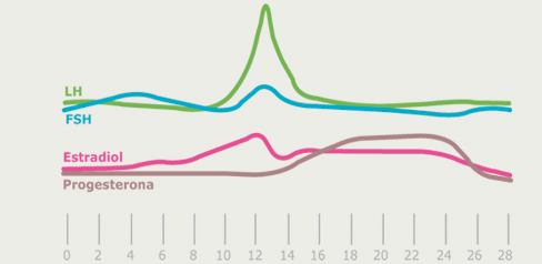
1. Check that women ovulate: if the woman menstruates every 28-30 days it is virtually certain that she has ovulatory cycles. Hormonal analysis: it allows us to know whether there is sufficient ovarian reserve. The blood sample test takes place on the third day of the cycle.

The “premature ovarian failure”(POF) is characterized by the existence of serum levels of follicle stimulating hormone (FSH) abnormally elevated in basal conditions (day 3 of cycle). Many of the patients will be within the group of women of reproductive age limit, so it is a key factor in the study of these patients.
It has been shown that serum levels of FSH predicted better ovarian response than age. These women have inadequate ovarian reserve, so the existence of a normal or abnormal ovarian reserve is crucial to the reproductive future of a woman and, in fact, the inadequate ovarian reserve is considered today as a cause of infertility.
2. Semen analysis: It allows us to verify that the semen is normal. We consider a normal sperm count when with a normal volume of ejaculate we have 20 million sperm per milliliter, with a useful mobility (progressive) 50% (rectilinear progress of 25% at least) and a top rate of 15% normal sperm.
Study of damage in the DNA of sperm:
The quality of chromatin in the sperm is being considered as a new parameter of seminal quality, possibly related to the fertility of the individual. There is evidence to suggest that the integrity of sperm DNA is associated with fertility of a couple and helps to predict the chances of pregnancy and their success. In natural conception, several studies indicate a significant impact of the damage DNA in sperm fertilization.
It should be noted that the fragmentation of the DNA does not seem to have a full correlation with any of the classic parameters of seminal quality (number, morphology and mobility). This means that men with normal sperm count could take a percentage of DNA fragmentation of its above normal, so it would compromise its ability to fertilize the oocyte.
Therefore, it is advisable to study the rate of sperm DNA fragmentation in order to take a more comprehensive picture of the seminal quality and, therefore, about the fertilization sperm ability.
3. To demonstrate that the female reproductive organs are normal:
Anatomical and functional uterine and tubal:
The uterus. The normal anatomy of the uterus can be easily checked by trasvaginal ultrasound, by hysterosalpingography (HSG) or by hysteroscopy. The ideal supplement Laparoscopy, allows the direct view of the uterine contour. The three methods will be complementary to the definitive diagnosis of the two most important diseases, the presence of myomas and uterine malformations.
The myomas can be easily identified with trasvaginal ultrasound, to assess their exact location and we can now even calculate quite accurately the relationship between myomas and uterine vessels with the use of color Doppler, which can be raised, on a rational acceptable basis, whether or not surgery is necessary. To make the differential diagnosis between myoma and other entities - such as polyps - hysteroscopy is essential, besides being a wonderful way to resolve surgically organic accidents present in the uterine cavity. A uterine malformation can also be easily detected by trasvaginal ultrasound.However, the accurate diagnosis will be done through laparoscopy because neither the HSG, or hysteroscopy, will be definitive for the differential diagnosis between a bicornual uterus and a septum. If the presence of a uterine wall is confirmed, the hysteroscopy will be definite in its final resection.
The tubes. The tubal patency can be determined only through the use of liquid substances added to the imaging techniques that identify the passage of the same from the uterine cavity to the outside of the abdominal cavity. Classically, we use the HSG and laparoscopy.
Today, we use different means of liquid next to the trasvaginal ultrasound, in what is called Sonohysterosalpingography. The advantage of the Sonohysterosalpingography is that it can be perform on an outpatient basis by the gynecologist, with few side effects and well tolerated by the patient.
The cervix. Both the HSG and ultrasound are helpful for assessing the extent of the cervical canal. Almost all the inefficiencies that we see are the result of curettage and not the cause of the abortions that led to them.
The assessment of the functional capacity of the cervix was traditionally achieved through the post-coital test that is not valid at present.

Patients area | First visit | Diagnostic tests | Psychological support | How to visit | Frequently asked questions | Oocyte donation | Sperm donation |
Documents | News | Legal notice | Credits: Deumonforte


